The History of Cataract Surgery
As part of Cataract Awareness month, we are continuing our series of articles on cataract surgery.
The origins of cataract surgery are embedded in antiquity, with the very first procedures performed in the fifth century BC¹. The procedure at the time used a technique called couching, and was performed only in extremely advanced cataracts.
Couching involved hitting the eye forcefully, causing the near-opaque lens to dislocate and fall into the back part of the eyeball, remaining inside the eye. Later, the same procedure was performed using a fine, sharp instrument which was used to break the delicate fibres holding the lens in place. The result was the same – the lens fell out of the line of sight, resulting in less cloudy, but completely unfocused vision. Whilst this was considered a great success initially, the damage caused to the eye by the technique itself, together with the inflammatory response inevitably caused by the retained lens material, often resulted in blindness.
The first true case of cataract surgery was performed by French surgeon Jacques Daviel in Paris in 1747. His procedure was more effective than couching, with an overall success rate of 50%². Surgery involved making a large incision around the cornea, through which the intact lens was extracted, leaving at least part of the lens capsule behind. This was a great improvement on couching, but was plagued with postoperative complications, including infection inside the eye, a result of the size of the wound and poor infection control. In the absence of sutures, the wound could not be sewn closed, so patients were confined to bed, with sandbags holding their heads in place until the wound healed. This meant that there was an even more serious potential complication, that of death secondary to stroke due to the long period of immobility.
Daviel’s surgical technique was known as extracapsular cataract extraction (ECCE), and continued to evolve over the following 200 years, swinging in and out of favour as other techniques were trialled. Perfecting removal of the lens whilst leaving the lens capsule intact was a critical stage of development, with the capsule acting to prevent any lens material falling into the back of the eye, avoiding the inflammatory response.
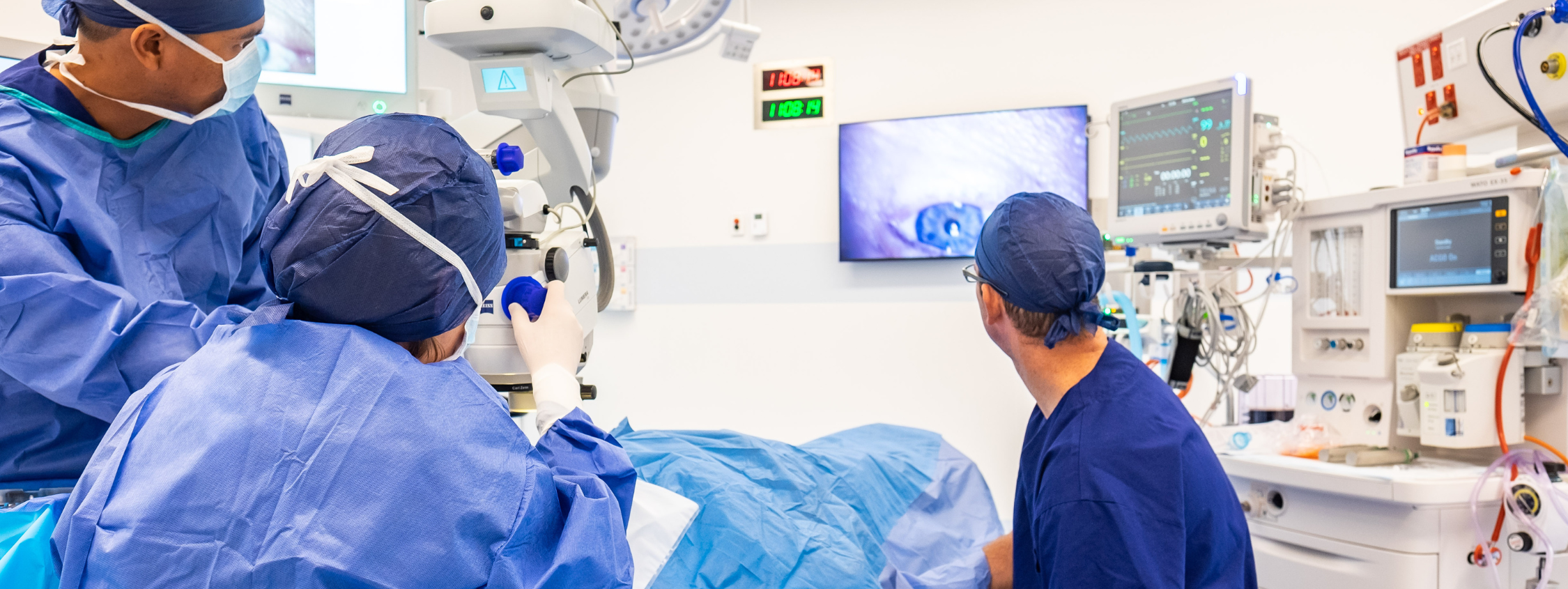
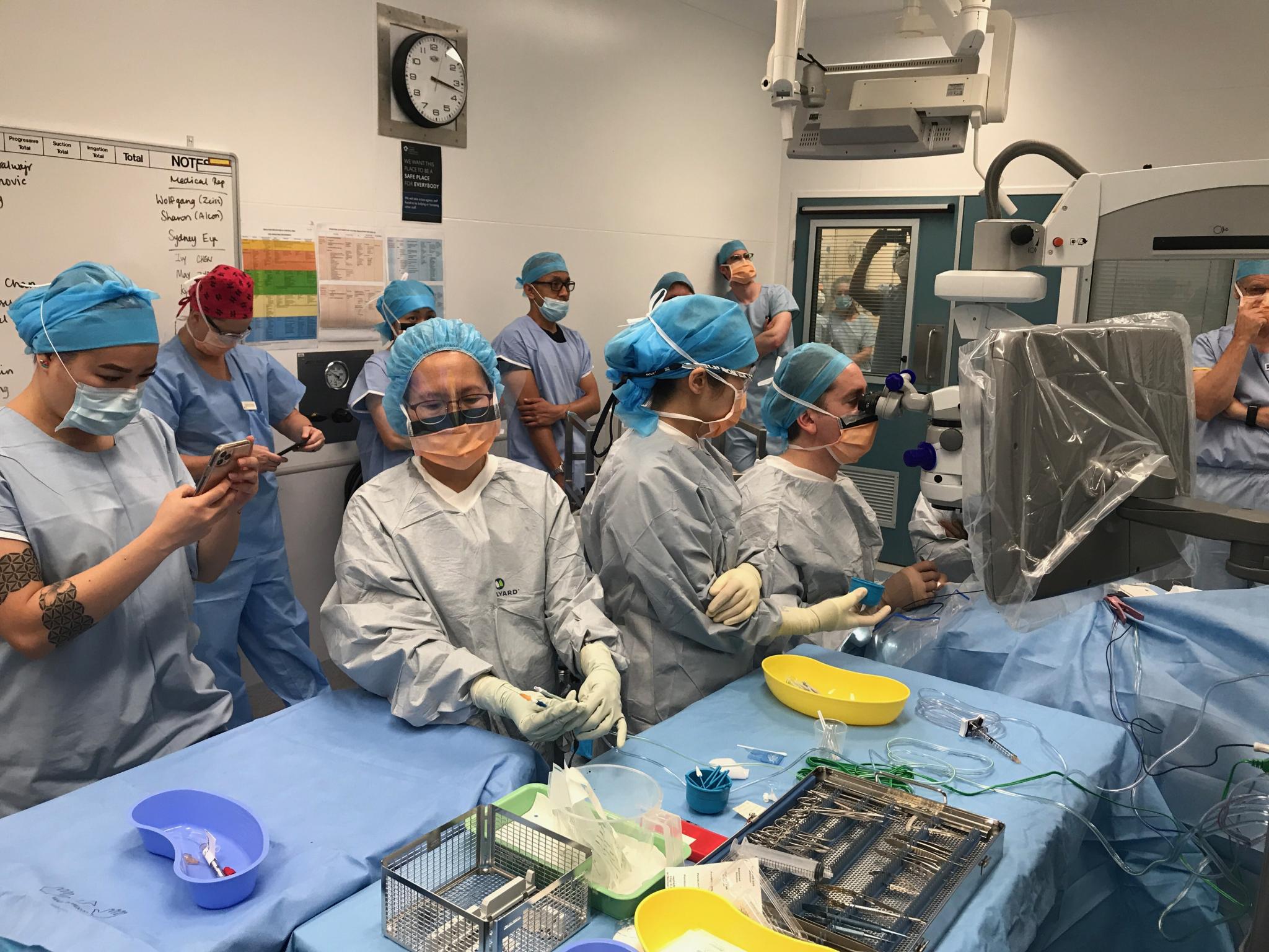
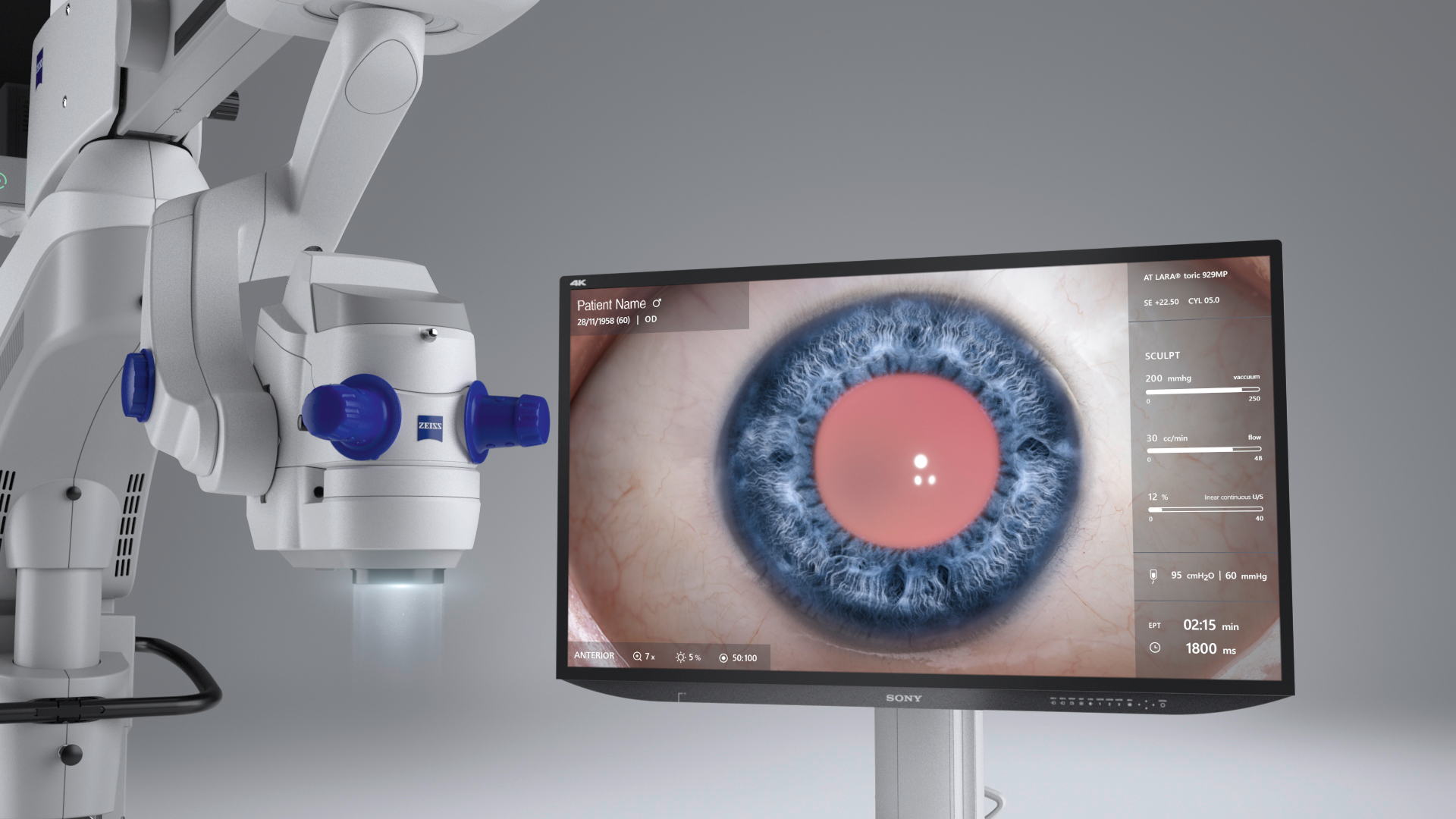
Modern cataract surgery evolved rapidly particularly during the second half of the 1900s, with the incision size decreasing which reduced the number of sutures required to seal the eye, as well as the visual distortion the residual scarring created. With the arrival of phacoemulsification surgery, developed by Charles Kelman in 1967, wound size decreased even more, and today can be less than 2mm. Phacoemulsification (from the Greek word phako – lens) is a process where a sophisticated ultrasound machine breaks the lens material up into tiny fragments, and removes it through a small incision, leaving the lens capsule intact and in place. The technique, though difficult to master, facilitates faster healing and reduced infection rates. In combination with improved cutting techniques, this small incision size allows the eye to self-seal and eliminates the need for sutures in most cases.
In parallel, the development of intraocular lenses has improved visual outcomes after surgery. The first intraocular lens was developed by British ophthalmologist, Dr Harold Ridley in the 1940s, and meant that once the cloudy cataract lens was removed, a clear man-made plastic lens could be inserted in its place. This allows clear, focused vision after surgery. Ridley’s discovery is a fascinating one. When treating Royal Air Force casualties during the Second World War, Ridley noticed that airmen with eye injuries due to shattered aircraft cockpit canopies often did not have ocular inflammation from the shards of Perspex left embedded in their eyes. This was in direct contrast to the shards from glass canopies,which triggered inflammatory rejection. Ridley correctly concluded from this that the acrylic plastic could be developed into a lens that could be left permanently inside the eye and went on to insert the first intraocular lens – itself made from Perspex. Since that time, the development of improved intraocular lens designs has radically transformed visual outcomes. In the early 1990s the foldable intraocular lens was developed, and could be inserted into the intact lens capsule, holding it securely in place. Accurate measurements are now taken to determine the optimum powered lens required to focus vision after surgery, with cataract surgery today capable of providing clear vision across a range of distances without the need for spectacles.
Cataract surgery is now one of the most commonly performed and successful elective procedures in Australia. 97% to 98% of all cases performed by an experienced surgeon are free of complications⁴ and Nexus Hospitals performs over 16,000 cataract extraction procedures annually. To locate one of our experienced cataract surgeons, please click here.
¹Cataract Surgery in Antiquity. American Academy of Ophthalmology. 2008
²Rucker CW. Cataract: A Historical Perspective. Invest Ophthalmol. 1965 Aug; 4:377-83
³Williams HP. Sir Harold Ridley’s vision. Br J Ophthalmol. 2001 Sep; 85(9):1022-3
⁴Clark A, et al. Whole Population Trends in Complications of Cataract Surgery Over 22 Years in Western Australia. Ophthalmology. 2011 Jun;118(6):1055-61.


